
Makeup
Eosinophilic Granuloma: Diagnosis, Treatment, and What to Know
Aug
A stubborn ache in the skull, jaw, rib, arm, or leg is usually blamed on an injury, an overenthusiastic workout, or the mysterious consequences of sleeping “wrong.” Occasionally, however, imaging reveals a small area where bone has been damaged. One rare possible explanation is eosinophilic granuloma.
Eosinophilic granuloma is generally considered the localized, usually bone-limited form of Langerhans cell histiocytosis, or LCH. It occurs when abnormal immune cells collect in tissue and create an inflammatory lesion. Although the terminology sounds like something produced by an allergy laboratory and a medical spelling bee working together, it is not the same as an ordinary eosinophilic or allergic disorder.
Most people with a single bone lesion have an excellent outlook, and some lesions heal with minimal treatment. Nevertheless, eosinophilic granuloma must be diagnosed carefully because infections, benign bone tumors, and malignant tumors can look similar on an X-ray. Doctors also need to determine whether the lesion is truly isolated or part of more widespread LCH.
What Is Eosinophilic Granuloma?
Eosinophilic granuloma is an older clinical name for a localized form of LCH, most commonly involving one bone. Modern medical classifications place LCH among clonal disorders of myeloid immune cells. The National Cancer Institute describes LCH as a rare blood cancer, while some patient resources still explain it as a cancer-like or inflammatory condition. The important practical point is that its behavior varies enormously: one lesion may resolve quietly, while multisystem disease requires specialist treatment.
Langerhans cells normally participate in immune surveillance. In LCH, abnormal cells accumulate with other inflammatory cells, including eosinophils, and may damage nearby bone or tissue. The presence of eosinophils helped inspire the name “eosinophilic granuloma,” but the abnormal Langerhans-type cells are the central players.
Genetic changes affecting the MAPK signaling pathway are found in many LCH lesions. These may include changes involving BRAF, MAP2K1, RAS, or ARAF. These mutations are usually acquired in the affected cells rather than inherited from a parent, so a diagnosis does not ordinarily mean that other family members will develop the condition.
Who Develops Eosinophilic Granuloma?
The condition is most frequently diagnosed in children, adolescents, and young adults, although it can appear at any age. Localized bone disease tends to occur in older children and adults compared with aggressive multisystem forms of LCH, which are more often seen in very young children.
Commonly affected bones include the skull, jaw, ribs, pelvis, spine, femur, and humerus. A lesion may occur in one bone, known as unifocal disease, or in several bones, known as multifocal bone disease. Bone is the most frequently involved organ system in LCH overall.
Symptoms of Eosinophilic Granuloma
Symptoms depend on the location and size of the lesion. Some lesions are discovered accidentally during imaging performed for an unrelated reason. Others behave like an annoyingly persistent bruise that never received the memo to go away.
Common Bone Symptoms
- Localized bone pain or tenderness
- Swelling or a noticeable lump
- Pain that persists despite rest
- Reduced movement near an affected joint
- Headache when the skull is involved
- Loose teeth, gum swelling, or jaw discomfort
- Back or neck pain when a vertebra is affected
- A fracture through weakened bone in uncommon cases
Skull lesions can appear as tender, slightly raised areas. Jaw involvement may resemble severe dental or periodontal disease. Vertebral lesions may weaken and flatten a vertebral body, producing a radiographic appearance called vertebra plana. Most spinal lesions do not injure the spinal cord, but weakness, numbness, difficulty walking, or loss of bladder control requires urgent evaluation.
Signs That May Suggest More Widespread LCH
A person with an apparently isolated bone lesion should tell the medical team about persistent rashes, excessive thirst, frequent urination, unexplained ear drainage, chronic cough, breathing problems, fever, fatigue, weight loss, or abdominal swelling. These symptoms do not prove multisystem LCH, but they may influence the diagnostic workup.
How Eosinophilic Granuloma Is Diagnosed
Medical History and Physical Examination
The clinician begins by asking when the pain or swelling started, whether an injury occurred, and whether symptoms are getting worse. The examination checks the affected bone, nearby joints, skin, lymph nodes, abdomen, neurological function, and other areas suggested by the patient’s symptoms.
Because eosinophilic granuloma is rare, the first suspected diagnosis may be infection, an ordinary bone cyst, a sports injury, or another bone tumor. That is not necessarily a mistake; several conditions share similar symptoms, and diagnosis is a process rather than a dramatic television reveal accompanied by ominous background music.
Imaging Tests
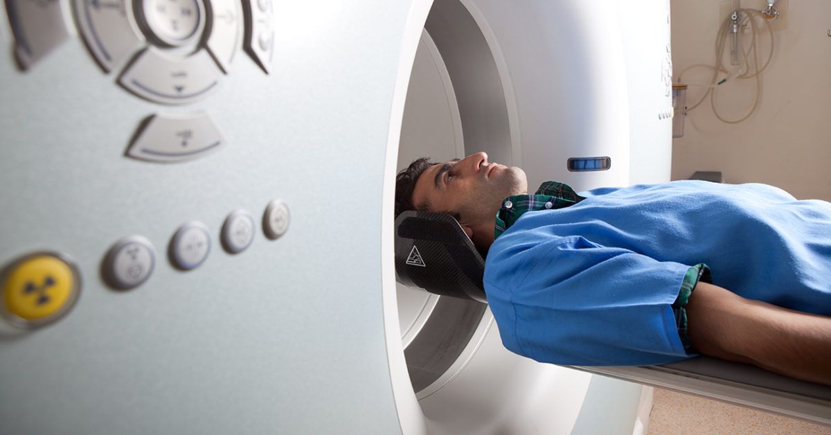
Plain X-rays often show an osteolytic lesion, meaning an area where bone has been broken down. Depending on its location, the lesion may look sharply defined or have less distinct margins. Computed tomography can show the bony detail more clearly, while magnetic resonance imaging is useful for evaluating bone marrow, nearby soft tissue, the spinal canal, or structures around the brain and eyes.
Additional imaging may be used to look for other lesions. The exact approach varies by age, symptoms, local protocols, and whether the patient has single-system or suspected multisystem disease. Imaging can identify suspicious areas, but it usually cannot prove eosinophilic granuloma on its own.
Biopsy: The Decisive Test
A tissue biopsy is generally required for a definitive diagnosis. A doctor may use a needle guided by CT imaging or perform a small surgical biopsy. A pathologist then examines the tissue and uses immunohistochemical stains to identify the abnormal cells.
LCH cells characteristically express CD1a and CD207, also called langerin. S100 is commonly positive but is less specific. Under electron microscopy, the cells may contain Birbeck granules, historically described as tennis-racket-shaped structures, although modern immunohistochemistry usually provides the necessary confirmation.
Tests to Determine the Extent of Disease
Once the biopsy confirms LCH, the medical team evaluates whether the condition involves only one bone, multiple bones, or other organs. Depending on the situation, the workup may include a complete blood count, liver and kidney tests, inflammatory markers, hormone testing, urine testing, chest imaging, abdominal ultrasound, skeletal imaging, MRI, or PET imaging. Molecular testing for mutations such as BRAF V600E may be useful in complicated, recurrent, multisystem, or treatment-resistant disease.
Conditions That Can Look Similar
Doctors must distinguish eosinophilic granuloma from osteomyelitis, Ewing sarcoma, osteosarcoma, leukemia or lymphoma involving bone, metastasis, bone cysts, osteoid osteoma, osteoblastoma, and other benign lesions. In the jaw, severe dental infection and periodontal disease may enter the discussion. Clinical findings, imaging, laboratory tests, and biopsy results are interpreted together rather than treated like separate contestants in a diagnostic game show.
Treatment for Eosinophilic Granuloma
Treatment is individualized according to the patient’s age, pain level, lesion location, fracture risk, number of lesions, organ involvement, and molecular findings. A single uncomplicated bone lesion is managed very differently from multisystem LCH.
Observation
Some small, asymptomatic, biopsy-confirmed lesions can be monitored because localized bone LCH may heal spontaneously. Observation does not mean forgetting that the lesion exists and hoping it develops good manners. It involves scheduled clinical examinations and repeat imaging to confirm healing and identify additional disease.
Curettage or Limited Surgery
Curettage involves opening the lesion and removing a limited amount of abnormal tissue. The biopsy itself may disrupt the lesion enough to stimulate healing. Complete radical removal is generally unnecessary for an isolated bone lesion and may cause avoidable structural damage, slower healing, or cosmetic problems.
Bone grafting or stabilization may be considered when a large lesion threatens a fracture. Spinal bracing, fixation, or rarely fusion may be needed if a vertebral lesion causes instability or neurological symptoms.
Intralesional Corticosteroid Injection
A corticosteroid may be injected directly into selected lesions after diagnosis. This local approach can reduce inflammation and pain while avoiding broader treatment. Its suitability depends on the location, accessibility, and structural risk of the lesion.
Systemic Therapy
Systemic treatment may be recommended for multiple bone lesions, lesions in certain skull-base or craniofacial locations, recurrent disease, soft-tissue extension, or involvement of organs outside the skeleton. Pediatric regimens may include vinblastine with prednisone, while cytarabine, cladribine, and other medicines are used in selected situations. Adult treatment plans may differ and should be directed by clinicians experienced in histiocytic disorders.
Targeted therapies that inhibit the MAPK pathway, including BRAF or MEK inhibitors, have become important options for some patients with mutation-positive, recurrent, or difficult-to-treat LCH. These medications are not routine treatment for every isolated eosinophilic granuloma.
Radiation Therapy
Low-dose radiation is now used selectively, especially in children, because many lesions respond to less invasive treatments and radiation can produce long-term effects. It may be considered for a painful or structurally dangerous lesion that cannot be treated adequately with surgery, local injection, or systemic therapy.
Recovery and Prognosis
The prognosis for a solitary bone lesion is generally excellent. Pain often improves before imaging looks completely normal, and visible bone repair may continue for months. A rim of new sclerosis around the lesion can be a sign of healing.
Recurrence is possible. The National Cancer Institute reports that reactivation risk is much lower in single-system unifocal bone disease than in multisystem LCH, but patients still require follow-up because new lesions or lasting complications can appear.
Long-term concerns depend on the original location. Possible complications include fracture, altered bone growth, spinal deformity, hearing or vision problems, dental damage, chronic pain, and hormone deficiencies. Craniofacial disease near the orbit, temporal bone, mastoid, sphenoid, or pituitary region deserves particular attention because it can be associated with endocrine or neurological complications.
When to Seek Medical Care
Arrange a medical evaluation for persistent focal bone pain, unexplained swelling, a growing lump, recurrent pain at night, loose teeth without an obvious dental cause, or pain that continues after an injury should have healed.
Seek urgent care for new weakness, numbness, trouble walking, loss of bladder or bowel control, severe breathing difficulty, sudden chest pain, a suspected fracture, rapidly worsening swelling, or changes in vision. These symptoms have many possible causes, but none deserve the “let’s see what happens next month” treatment plan.
Experiences During Diagnosis, Treatment, and Follow-Up
The following examples are educational composite scenarios based on commonly reported patient journeys. They do not describe one identifiable patient and should not replace individualized medical advice.
The Child With a Mysterious Skull Bump
A common journey begins with a parent noticing a tender bump on a child’s scalp. The child may remember bumping their head during sports, so the family initially expects the swelling to disappear. When it remains painful or grows, the pediatrician orders an X-ray or CT scan. The phrase “bone lesion” appears in the radiology report, and the family’s stress level immediately launches into orbit.
The next several days may involve an orthopedic surgeon, neurosurgeon, oncologist, or interventional radiologist. Families often say that the uncertainty before biopsy is harder than the procedure itself. Eosinophilic granuloma is rare enough that nobody expected to meet it, yet common enough within pediatric bone-lesion clinics that specialists usually have a clear diagnostic plan.
After a biopsy confirms a solitary lesion, the treatment may be limited to curettage or observation. The child may return to school quickly but temporarily avoid contact sports. Follow-up scans can remain abnormal even when pain is gone, which may worry parents until the clinician explains that bones remodel on their own leisurely schedule.
The Adult Whose “Pulled Muscle” Would Not Quit
An adult may develop persistent rib, shoulder, hip, or back pain after exercise. At first, the discomfort behaves enough like a strain that rest, ice, and over-the-counter medication seem reasonable. Weeks later, the pain is still submitting daily attendance reports.
Imaging reveals a small lytic lesion. Because eosinophilic granuloma is uncommon in adults, the diagnostic conversation may include infection, metastasis, multiple myeloma, or a primary bone tumor. Hearing those possibilities can be frightening, even when the clinician emphasizes that they are items on a differential diagnosis rather than confirmed conclusions.
Biopsy can bring both relief and a new vocabulary lesson: Langerhans cells, CD1a, langerin, MAPK pathway. If staging shows only one lesion, treatment may be local and recovery relatively straightforward. The emotional recovery may take longer. Many adults remain alert to every ache afterward, wondering whether it is a new lesion or simply the ordinary creaking of a human skeleton. A clear follow-up schedule helps keep reasonable monitoring from turning into full-time symptom surveillance.
Living With Follow-Up
Follow-up appointments are an important part of the experience even after successful treatment. Patients or parents may keep notes about pain, swelling, thirst, urination, skin changes, headaches, hearing, and activity levels. Bringing these observations to visits is helpful; performing hourly inspections with a flashlight generally is not.
Questions about sports, school, work, dental treatment, travel, and pain medication are normal. A patient with a healing leg lesion may need temporary activity restrictions, while someone with a small skull lesion may have few limitations. The plan should reflect lesion location and structural strength rather than the diagnosis alone.
Rare diseases can also feel socially isolating. Friends may assume that “benign” means trivial, while the words “neoplasm,” “chemotherapy,” or “oncology clinic” sound anything but trivial. Both reactions miss the nuance. Localized eosinophilic granuloma is usually highly treatable, yet the diagnostic process and possibility of recurrence can still create real anxiety. Support from an experienced clinical team, family members, counselors, or rare-disease communities can make the uncertainty easier to manage.
Conclusion
Eosinophilic granuloma is the localized form of Langerhans cell histiocytosis most often found in bone. It may cause pain, swelling, headaches, dental symptoms, or an incidental abnormality on imaging. Because its appearance can resemble infection and other bone tumors, diagnosis generally requires a biopsy with characteristic immunohistochemical findings.
Many solitary lesions heal after observation, limited curettage, or a local corticosteroid injection. Multiple lesions, high-risk locations, recurrence, or organ involvement may require systemic therapy and multidisciplinary care. Most patients with isolated bone disease do very well, but appropriate staging and long-term follow-up remain important.
Medical note: This article provides general educational information and is not a substitute for diagnosis or treatment by a qualified healthcare professional. Anyone with a suspected bone lesion or confirmed LCH should receive an individualized evaluation from an appropriate specialist.

