
Makeup
DVT Ultrasound: Procedure, Accuracy, Next Steps, and More
Jul
A suddenly swollen, sore, or unusually warm leg can inspire several theories, ranging from “I overdid leg day” to “my body has launched a surprise medical subplot.” One important possibility is deep vein thrombosis, or DVTa blood clot that develops in a deep vein, most commonly in the leg. Because part of a clot can travel to the lungs and cause a pulmonary embolism, suspected DVT deserves prompt medical evaluation rather than a wait-and-see experiment.
A DVT ultrasound is usually the first imaging test used to investigate that possibility. It is noninvasive, does not expose you to ionizing radiation, and can show whether a vein collapses normally under gentle pressure and whether blood is flowing through it as expected. This guide explains how the procedure works, how accurate it is, what the results may mean, and what commonly happens next.
What Is a DVT Ultrasound?
A DVT ultrasound is an imaging examination of the deep veins, usually in one or both legs. You may also hear it called a venous ultrasound, duplex ultrasound, Doppler ultrasound, or compression ultrasonography. These names describe related parts of the same basic investigation.
The test uses high-frequency sound waves to create real-time images of veins beneath the skin. Standard grayscale imaging shows the vein’s structure, while Doppler technology evaluates the direction and speed of blood flow. During a compression study, the sonographer gently presses the ultrasound probe against selected sections of the vein. A healthy vein generally flattens under pressure. A vein containing a clot may not compress completely.
What areas are examined?
Most examinations focus on the veins running from the groin through the thigh, behind the knee, and sometimes into the calf. If symptoms involve an armpossibly after placement of a catheter, pacemaker lead, or another medical devicean upper-extremity venous ultrasound may be performed instead. Ultrasound duplex Doppler is considered an appropriate first imaging study for suspected DVT in either the lower or upper extremity.
Why Might a Healthcare Professional Order the Test?
A clinician may order a DVT ultrasound when symptoms, medical history, or recent circumstances suggest that a blood clot is possible. Common warning signs include:
- Swelling in one leg or arm
- Pain, aching, cramping, or tenderness without a clear injury
- Skin that feels warmer than the surrounding area
- Red, bluish, or otherwise discolored skin
- Newly prominent surface veins
- Unexplained limb heaviness or tightness
Not everyone with DVT has obvious symptoms, and those who do may have symptoms resembling a muscle injury, cellulitis, a ruptured Baker’s cyst, lymphedema, or chronic venous disease. That is why diagnosing DVT by appearance alone is unreliable. The leg cannot simply be asked whether it is harboring a clot; unfortunately, it remains committed to mystery.
Risk factors that may raise suspicion
Recent surgery, hospitalization, major injury, prolonged immobility, pregnancy, estrogen-containing medication, active cancer, previous DVT, inherited clotting conditions, and long periods of sitting can increase the likelihood of venous thrombosis. Risk factors do not prove that a clot exists, but they help clinicians estimate the pretest probability before choosing blood tests or imaging.
How DVT Is Evaluated Before Ultrasound
Healthcare professionals commonly begin with a medical history and physical examination. They may use a validated clinical prediction rule, such as the Wells score, to classify DVT as more or less likely. The score considers findings such as leg swelling, tenderness along deep veins, recent immobilization, cancer, previous DVT, and whether another diagnosis seems equally plausible.
For a patient with a low clinical probability, a negative D-dimer blood test may be enough to rule out DVT without ultrasound. D-dimer is a fragment produced when the body breaks down a clot. It is sensitive but not specific: infection, inflammation, surgery, pregnancy, aging, and several other situations can increase it. A positive result therefore does not announce, “Congratulations, you definitely have a DVT.” It means additional testing may be needed. Patients with a high pretest probability generally require imaging even when a D-dimer result is negative.
Types of DVT Ultrasound
Compression ultrasound
Compression is the most important feature of many DVT examinations. The sonographer views a vein in cross-section and presses down with the transducer. Failure of the vein walls to meet completely is a major sign of thrombosis.
Duplex and color Doppler ultrasound
Duplex imaging combines a structural image with Doppler measurements. Color Doppler may display blood movement using colors, while spectral Doppler creates a waveform representing flow. These tools help identify partial obstruction, absent flow, or changes suggesting a blockage farther up the venous system.
Proximal ultrasound
A proximal examination concentrates on the major veins of the thigh and the area behind the knee. Clots in these veins are especially important because proximal DVT has a greater association with pulmonary embolism than an isolated calf clot. A negative proximal scan may need to be repeated when clinical suspicion remains significant.
Whole-leg ultrasound
A whole-leg study includes the calf veins as well as the proximal veins. A technically adequate negative whole-leg ultrasound can often rule out lower-extremity DVT in a single visit. The trade-off is that it may identify small isolated calf clots whose ideal management depends on symptoms, extension risk, bleeding risk, and the patient’s overall circumstances.
How to Prepare for a DVT Ultrasound
Preparation is generally refreshingly uneventful. You usually do not need to fast, receive sedation, or stop regular medication unless a healthcare professional gives you specific instructions. Wear loose clothing that allows access from the groin to the ankle. You may be asked to change into a gown and remove compression garments, bandages, or jewelry near the examination area.
Bring a medication list and be prepared to describe when the symptoms began, where they are located, and whether you have had a previous blood clot. Old ultrasound reports can be particularly useful in someone with prior DVT because residual vein changes may persist after treatment. Comparison can help a radiologist determine whether an abnormality is old, new, or impossible to classify with confidence.
What Happens During the Procedure?
- Positioning: You will usually lie on an examination table with the leg slightly turned outward. The sonographer may raise the head of the table or ask you to sit or stand for part of the study.
- Gel application: A clear, water-based gel is placed on the skin. The gel helps sound waves travel between the probe and your body.
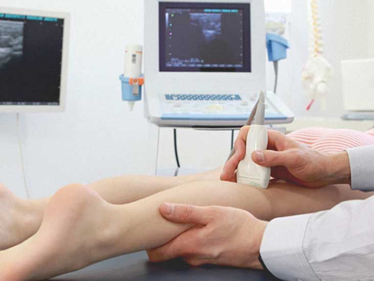
- Scanning: The sonographer moves a handheld transducer along the leg, following the course of the deep veins.
- Compression: The probe is pressed at intervals to see whether each vein collapses completely.
- Flow testing: Doppler measurements evaluate blood flow. The sonographer may gently squeeze the calf or ask you to breathe in a particular way to observe changes in venous flow.
- Image review: Images and measurements are saved for interpretation by a radiologist, vascular specialist, or another qualified clinician.
The scan often takes approximately 20 to 45 minutes, although bilateral studies, difficult anatomy, severe swelling, prior DVT, or a suspected clot in the pelvis can make the process longer. The examination does not normally require needles, contrast dye, or radiation.
Does a DVT ultrasound hurt?
The sound waves do not hurt. The compression portion can be uncomfortable when the leg is already tender or swollen because the sonographer must apply enough pressure to test the vein properly. Tell the sonographer when an area is painful. Discomfort is usually brief, and there is no award for pretending your calf has no nerve endings.
How Accurate Is a DVT Ultrasound?
Ultrasound is highly effective for detecting DVT in the larger proximal veins, but it is less sensitive for small clots in the calf. An evidence summary used by the American College of Radiology reported approximately 94% sensitivity for proximal DVT, 64% sensitivity for distal DVT, and 94% specificity. Sensitivity describes how often a test detects disease when it is present; specificity describes how often it correctly identifies people who do not have the disease.
Those figures are useful benchmarks, not a guarantee for every patient. Accuracy depends on the scanning protocol, the location and age of the clot, image quality, the experience of the examiner, and the patient’s anatomy. A scan may be more difficult in the presence of major edema, obesity, severe pain, a cast, open wounds, or veins located deep in the pelvis.
Why calf and pelvic clots are harder to detect
Calf veins are smaller and more numerous than proximal veins. Some occur in pairs, and their anatomy can be challenging to follow. Pelvic veins may be partly hidden by bone, bowel gas, and body depth. If symptoms strongly suggest an iliac or pelvic clot but the leg ultrasound is inconclusive, clinicians may consider CT venography, MR venography, or another imaging strategy.
Can an old clot affect the result?
Yes. A previous DVT can leave thickened vein walls, scarring, incomplete compressibility, or persistent material inside the vein. These chronic changes may resemble recurrent thrombosis. Prior imaging is therefore valuable. A new noncompressible venous segment or a meaningful increase in clot diameter compared with an earlier study can support a diagnosis of recurrence.
Understanding DVT Ultrasound Results
Positive result
A positive result means the scan found evidence of thrombosis. The report should identify the affected vein, the extent of the clot, whether it is proximal or distal, and whether it appears acute, chronic, or indeterminate in age. A clot in the femoral, popliteal, common femoral, or iliac system is generally considered proximal. Treatment often begins promptly to prevent clot growth, recurrence, and pulmonary embolism.
Negative result
A negative result means no DVT was seen in the veins adequately examined. Whether that ends the investigation depends on the type of ultrasound and the original clinical probability. A negative whole-leg study may be sufficient. After a negative proximal-only scan, selected patients with continuing symptoms, a positive D-dimer, or moderate-to-high suspicion may need a repeat compression ultrasound about one week later. The repeat scan checks whether an initially undetected calf clot has extended into a proximal vein.
Inconclusive or limited result
A study can be labeled limited when parts of the venous system cannot be seen or compressed adequately. The next step may be repeat ultrasound, examination at a specialized vascular laboratory, or CT or MR venography. An inconclusive scan should not be interpreted as automatically negative.
Chronic or age-indeterminate findings
Some reports describe chronic post-thrombotic changes or an abnormality of indeterminate age. Clinicians combine the images with symptoms, previous scans, laboratory results, and treatment history. This is especially important because anticoagulation has real benefits but also increases bleeding risk.
What Happens After a Positive DVT Ultrasound?
The main treatment for most confirmed DVTs is anticoagulation. These medications are commonly called blood thinners, although they do not literally turn blood into a fashionable beverage. They reduce the blood’s ability to form additional clot, helping prevent the existing clot from enlarging while the body gradually breaks it down.
The specific medication and treatment duration depend on clot location, kidney and liver function, pregnancy status, cancer, bleeding risk, medication interactions, cost, and whether the DVT was provoked by a temporary risk factor. Many patients receive at least three months of treatment, but the appropriate duration must be individualized.
When more intensive treatment may be considered
Selected patients with extensive iliac or common femoral DVT, severe symptoms, threatened limb circulation, or a high risk of long-term complications may be evaluated for catheter-based clot removal or thrombolytic therapy. These treatments are not routine for every DVT because they can cause serious bleeding. A vena cava filter is generally reserved for selected situations, such as when anticoagulation cannot be given safely.
Managing isolated calf DVT
Not every isolated distal DVT is managed identically. Anticoagulation may be recommended when symptoms are substantial or the clot has features associated with extension. In selected patients with a lower extension risk or a high bleeding risk, clinicians may choose serial ultrasoundoften weekly for about two weeksto watch for movement into proximal veins.
What If the Ultrasound Is Negative but Symptoms Continue?
Contact the treating healthcare professional if swelling, pain, warmth, or discoloration persists or worsens. Possible next steps include repeat ultrasound, a whole-leg study, alternative imaging, or evaluation for another condition. Muscle injury, cellulitis, a Baker’s cyst, superficial thrombophlebitis, chronic venous insufficiency, lymphedema, joint inflammation, and other problems can resemble DVT.
A negative scan should not overrule rapidly changing symptoms. Clinical probability and the technical quality of the examination still matter. In other words, “negative” is important information, but it is not always the final scene.
When to Seek Emergency Care
Call 911 or seek emergency care immediately for symptoms that may indicate pulmonary embolism, including:
- Sudden shortness of breath
- Chest pain, especially pain that worsens with a deep breath or cough
- Coughing up blood
- A rapid or irregular heartbeat
- Lightheadedness, fainting, confusion, or severe weakness
- New difficulty breathing alongside leg swelling or pain
A pulmonary embolism can occur when part of a deep vein clot travels to the lungs and blocks blood flow. It requires immediate medical attention. Do not drive yourself when symptoms are severe or fainting is possible.
Questions to Ask After the Scan
- Was the entire leg examined or only the proximal veins?
- Were any areas difficult to visualize?
- Is the result positive, negative, chronic, or inconclusive?
- Which vein is affected, and is the clot proximal or distal?
- Do I need a repeat ultrasound, and on what date?
- Should I start or continue anticoagulant medication?
- Which bleeding symptoms require urgent medical attention?
- Are there restrictions on walking, exercise, work, or travel?
- Could another condition explain my symptoms?
- Should previous ultrasound images be obtained for comparison?
Write down the answers or request a copy of the radiology report. Medical vocabulary has an impressive ability to disappear from memory approximately seven seconds after an appointment ends.
A Realistic DVT Ultrasound Experience: What the Day May Feel Like
The following is a composite description of experiences commonly associated with a DVT evaluation. It is not the story of a specific patient and should not replace individual medical advice.
The experience often begins with uncertainty rather than drama. Perhaps one calf looks slightly larger than the other after a long flight. Maybe the leg feels tight, warm, and oddly heavy. The person may spend several hours debating whether it is a pulled muscle, dehydration, an enthusiastic reaction to walking through an airport, or something more serious. Once a clinician hears about unilateral swelling and a recent period of immobility, the conversation usually becomes more focused.
At an urgent care center or emergency department, a nurse may record vital signs and ask about chest pain, breathing difficulty, recent surgery, pregnancy, medication, cancer treatment, and previous clots. The clinician examines both legs, measures swelling, and presses along areas where the deep veins run. A D-dimer blood test may be ordered, although some patients proceed directly to ultrasound because their clinical probability is high enough that imaging is required regardless of the blood result.
The ultrasound room is usually dim and quiet. The patient lies on a table while the sonographer applies gel near the groin. That location can be surprising to someone who assumed the scan would focus only on the sore calf, but major deep veins begin higher in the leg. The transducer moves slowly downward. Every few inches, the sonographer presses firmly to test whether a vein compresses. Most of the exam feels like mild pressure. Over the most tender area, however, the pressure can produce a sharp “yes, that is definitely the spot” reaction.
The sonographer may squeeze the calf while watching the screen, ask the patient to hold a breath briefly, or reposition the leg. The patient may attempt to decode the grayscale images and colored Doppler signals, an activity roughly as successful as interpreting satellite communications from another galaxy. Sonographers often cannot discuss a diagnosis during the scan because the images must be reviewed according to the facility’s process.
If the ultrasound is negative and the clinical suspicion is low, the patient may leave with advice about another likely cause of the symptoms. When suspicion remains elevated, the discharge instructions may include a specific date for repeat imaging. That detail matters. “Repeat in one week” should become an actual appointment, not a note destined to live forever beneath old grocery receipts.
If the scan is positive, the next conversation may happen quickly. A clinician explains where the clot is located, whether it is proximal or confined to the calf, and whether treatment should start immediately. Many stable patients can be treated without hospital admission, but this depends on bleeding risk, symptom severity, other medical conditions, access to medication, and the possibility of pulmonary embolism.
The emotional response can range from relief that the cause was found to fear about the word “clot.” It helps to remember that DVT is treatable and that prompt diagnosis substantially changes the situation. Before leaving, the patient should understand the medication plan, warning signs of bleeding, symptoms of pulmonary embolism, follow-up arrangements, and whom to contact with questions. A five-minute written checklist is often more useful than trying to preserve an entire medical conversation using adrenaline alone.
Conclusion
A DVT ultrasound is a fast, noninvasive, and highly useful test for investigating suspected blood clots in the deep veins. Its strongest performance is in the larger proximal leg veins, while calf and pelvic clots can be more challenging. The test works by examining vein structure, compressibility, and blood flowwithout radiation or routine contrast injection.
A positive scan usually leads to prompt treatment planning. A negative scan may end the evaluation when clinical probability is low or a complete whole-leg examination is negative. When suspicion remains high, repeat ultrasound or alternative imaging may still be necessary. Most importantly, sudden shortness of breath, chest pain, coughing blood, or fainting should be treated as an emergency rather than an invitation to conduct one more internet search.

