Hair Care
Melanoma Stages: Diagnostic Testing and Treatment Options
Jun
Melanoma is the skin cancer that likes to act like the quiet guest at the partyeasy to overlook at first, but capable of causing serious trouble if ignored. It begins in melanocytes, the pigment-making cells that give skin its color, and it can develop in an existing mole or appear as a brand-new spot. While melanoma is less common than basal cell and squamous cell skin cancers, it is more likely to spread if it is not found and treated early.
The good news? Melanoma is often highly treatable when caught early. The even better news? Doctors now have more tools than ever to diagnose, stage, and treat itfrom precise biopsies and sentinel lymph node testing to immunotherapy, targeted therapy, and advanced cellular treatments for certain metastatic cases. In other words, melanoma care has moved far beyond “remove it and hope.” Today, staging helps build a personalized treatment plan.
This guide explains melanoma stages, diagnostic testing, and treatment options in clear American Englishno medical fog machine required.
What Does “Melanoma Stage” Mean?
Melanoma staging describes how far the cancer has grown or spread. Doctors use staging to estimate risk, choose treatment, plan follow-up care, and decide whether additional testing is needed. The main system is called TNM:
- T stands for tumor: how thick the melanoma is, whether it is ulcerated, and how deeply it has grown.
- N stands for nodes: whether melanoma cells have reached nearby lymph nodes or nearby skin/lymphatic channels.
- M stands for metastasis: whether the melanoma has spread to distant organs such as the lungs, liver, brain, bones, or distant skin.
A melanoma’s stage is not just a label. It is the GPS for treatment. Stage 0 may need only local surgery, while stage III or IV may involve surgery plus immunotherapy, targeted therapy, radiation, clinical trials, or a combination approach.
The Main Melanoma Stages Explained
Stage 0 Melanoma: Melanoma in Situ
Stage 0 melanoma means abnormal melanocytes are limited to the top layer of skin, the epidermis. “In situ” is doctor-speak for “still in place,” which is one of the few medical phrases that sounds fancy but is actually comforting.
Common treatment: surgical excision, removing the melanoma with a margin of normal-looking skin. Because it has not invaded deeper layers, stage 0 melanoma usually does not require lymph node testing or systemic treatment.
Stage I Melanoma: Early Invasive Melanoma
Stage I melanoma has grown into the skin but is still relatively thin and has not spread to lymph nodes or distant organs. The pathology report may mention Breslow thickness, which measures how deep the melanoma is in millimeters. A thinner melanoma generally has a lower risk of spread.
Common treatment: wide local excision. In selected cases, especially when the melanoma is thicker or has higher-risk features such as ulceration, doctors may discuss sentinel lymph node biopsy.
Stage II Melanoma: Thicker but No Detected Spread
Stage II melanoma is thicker and may be ulcerated, but testing does not show spread to lymph nodes or distant sites. This stage can feel confusing because the cancer is still “local,” yet some stage II melanomas carry a meaningful risk of recurrence.
Common treatment: wide local excision. Sentinel lymph node biopsy is often discussed because lymph node status can change the stage and treatment plan. For higher-risk stage IIB and IIC melanoma, adjuvant immunotherapy may be considered after surgery to reduce the risk of recurrence.
Stage III Melanoma: Spread to Nearby Nodes or Skin
Stage III melanoma means cancer has spread to nearby lymph nodes, nearby skin, or lymphatic channels between the original tumor and nearby nodes. This does not automatically mean the situation is hopeless, but it does mean the treatment plan usually becomes more aggressive.
Common treatment: surgery when the melanoma can be removed, possible lymph node management, and often systemic therapy. Options may include immunotherapy, targeted therapy for tumors with a BRAF mutation, or neoadjuvant therapy given before surgery in selected cases.
Stage IV Melanoma: Distant Metastatic Melanoma
Stage IV melanoma means the cancer has spread to distant organs or distant skin/lymph nodes. Common metastatic sites include the lungs, liver, brain, bones, and distant areas of skin. Treatment usually focuses on controlling disease, shrinking tumors, improving survival, and protecting quality of life.
Common treatment: immunotherapy, targeted therapy if the tumor has a BRAF V600 mutation, radiation therapy for specific symptoms or brain metastases, surgery in selected cases, clinical trials, and in some previously treated cases, tumor-infiltrating lymphocyte therapy.
How Melanoma Is Diagnosed
Skin Exam and the ABCDE Rule
Diagnosis often starts with a skin exam. Dermatologists look for suspicious spots using visual inspection and sometimes dermoscopy, a handheld tool that magnifies skin structures. The ABCDE rule is a helpful memory trick:
- A: Asymmetry one half does not match the other.
- B: Border edges look uneven, notched, blurred, or ragged.
- C: Color multiple colors or unusual shades appear.
- D: Diameter larger than about 6 millimeters, though melanomas can be smaller.
- E: Evolving changing size, shape, color, sensation, or behavior.
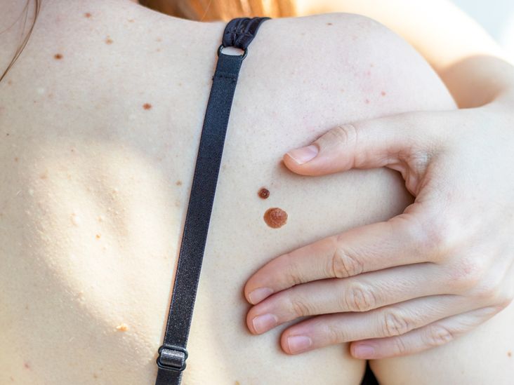
One warning sign deserves its own spotlight: the “ugly duckling” mole. If one spot looks different from the person’s other moles, it deserves attention. Skin is allowed to have freckles, dots, and personality. A suspicious mole, however, should not be freelancing as modern art.
Biopsy: The Test That Confirms Melanoma
A biopsy is the key test for diagnosing melanoma. When possible, doctors often remove the entire suspicious lesion with a narrow margin so a pathologist can examine it under a microscope. The pathology report may include:
- Breslow thickness
- Ulceration status
- Mitotic rate or growth activity
- Margins
- Subtype of melanoma
- Evidence of lymphovascular invasion or other high-risk features
These details help determine the melanoma stage and whether additional procedures are recommended.
Sentinel Lymph Node Biopsy
A sentinel lymph node biopsy checks the first lymph node or nodes where melanoma would most likely spread. A tracer and/or dye helps the surgeon identify these nodes, which are removed and examined by a pathologist.
This test is not used for every melanoma. It is typically considered when the melanoma has enough thickness or high-risk features to make lymph node spread more likely. A positive sentinel node can change the melanoma from stage I or II to stage III and may open the door to additional treatment options.
Imaging Tests
Imaging is not always needed for very early melanoma. For higher-stage disease, symptoms, abnormal exam findings, or suspected spread, doctors may order CT scans, PET/CT scans, MRI scans, or brain MRI. Imaging helps look for melanoma in lymph nodes or distant organs.
Think of imaging as a searchlight. Doctors do not shine it everywhere for every small mole, but when the stage or symptoms suggest risk, it becomes a powerful tool.
Blood Tests and LDH
Blood tests do not diagnose melanoma. However, they may be used before or during treatment, especially for advanced melanoma. Lactate dehydrogenase, often called LDH, may be checked in metastatic melanoma because a high LDH level can be linked with more difficult-to-treat disease.
Genetic and Molecular Testing
For stage III or stage IV melanoma, tumor testing may look for mutations such as BRAF V600. This matters because BRAF-positive melanoma may respond to targeted therapy using a BRAF inhibitor combined with a MEK inhibitor. Molecular testing helps doctors choose treatment based on the tumor’s biologynot just its address on the skin.
Treatment Options by Melanoma Stage
Surgery: The Foundation of Early Treatment
Surgery is the main treatment for stage 0, stage I, and many stage II melanomas. The surgeon removes the melanoma plus a safety margin of normal skin. The exact margin depends on melanoma thickness and location. In cosmetically sensitive areas, such as the face, doctors balance complete removal with preserving appearance and function.
For many early melanomas, surgery may be the only treatment needed. That is why early diagnosis matters so much: catching melanoma early can keep the treatment plan simpler and less dramatic than a hospital TV show.
Immunotherapy
Immunotherapy helps the immune system recognize and attack melanoma cells. Checkpoint inhibitors are a major category and include drugs that target PD-1, CTLA-4, or LAG-3 pathways. These medicines may be used after surgery for higher-risk melanoma or as treatment for unresectable or metastatic melanoma.
Examples include pembrolizumab, nivolumab, ipilimumab, and nivolumab combined with relatlimab. These treatments can be powerful, but they may also cause immune-related side effects because an activated immune system can sometimes attack normal organs. Patients should report symptoms such as severe diarrhea, worsening cough, shortness of breath, unusual fatigue, rash, yellowing skin, or severe headaches promptly.
Targeted Therapy
Targeted therapy is used for melanomas with certain mutations, especially BRAF V600 mutations. BRAF inhibitors are usually paired with MEK inhibitors because the combination works better than using a BRAF inhibitor alone and can help delay resistance.
Common combinations include dabrafenib plus trametinib, vemurafenib plus cobimetinib, and encorafenib plus binimetinib. These medicines are often taken by mouth, which sounds convenient until you remember they still require careful monitoring. “Pill” does not mean “casual.”
Radiation Therapy
Radiation therapy uses high-energy beams to target cancer cells. It is not usually the main treatment for early melanoma, but it can help in selected cases, such as treating brain metastases, relieving bone pain, controlling symptoms, or reducing recurrence risk in certain lymph node areas.
Chemotherapy
Chemotherapy is used much less often than it was in the past because immunotherapy and targeted therapy have changed melanoma care. Still, it may be considered in certain situations when other options are not appropriate or have stopped working.
Cellular Therapy and Clinical Trials
For some adults with unresectable or metastatic melanoma that has already been treated with other therapies, tumor-infiltrating lymphocyte therapy may be an option. This approach uses immune cells taken from the tumor, expanded in a lab, and returned to the patient. Clinical trials may also offer access to promising combinations, new drugs, vaccines, or personalized therapies.
Patients should not think of clinical trials as a “last resort closet.” Many trials are carefully designed treatment options that may be available at different points in care.
How Doctors Choose a Treatment Plan
Melanoma treatment depends on more than stage alone. Doctors also consider the melanoma’s thickness, ulceration, lymph node status, mutation profile, location, whether surgery can remove all visible disease, the patient’s age, other health conditions, immune system function, and personal goals.
For example, two people with stage III melanoma may have different plans. One may have surgery followed by adjuvant immunotherapy. Another may receive neoadjuvant immunotherapy before surgery because the lymph node disease is bulky but removable. A third may receive targeted therapy if the tumor has a BRAF mutation and that approach fits the clinical picture.
Follow-Up After Melanoma Treatment
Melanoma follow-up is not a “see you never” situation. Regular skin exams and physical exams are important because melanoma can recur, and people who have had melanoma are at increased risk for another melanoma. Follow-up schedules vary by stage and risk level, but visits are often more frequent in the first few years after treatment.
Patients may also be taught how to perform skin self-exams. A practical approach is to check from scalp to soles once a month, using mirrors or a partner for hard-to-see areas. Take photos of moles that are being monitored. Your camera roll may include pets, food, and one suspicious freckle named Gary. That is acceptable.
Prevention Still Matters
Not every melanoma is preventable, but reducing ultraviolet exposure helps lower risk. Good habits include using broad-spectrum sunscreen, wearing protective clothing, seeking shade, avoiding indoor tanning beds, and being extra careful around reflective surfaces like water, sand, and snow.
Sunscreen is not a force field. It works best when applied generously and reapplied, especially after swimming or sweating. Hats, sunglasses, and shade are not old-fashioned; they are your skin’s security team.
Real-Life Experiences and Practical Lessons About Melanoma Stages
One of the most common experiences people describe after a melanoma diagnosis is surprise. The spot often “didn’t look that bad.” It may have been small, flat, or only slightly darker than the surrounding skin. Some people notice itching or bleeding, but many do not. That is why the evolving part of ABCDE is so important. A mole does not need to look like a movie villain to deserve a biopsy.
Another common experience is confusion after receiving the pathology report. Terms like Breslow thickness, ulceration, margins, mitotic rate, and sentinel lymph node biopsy can make a patient feel as if they accidentally enrolled in medical school. A helpful strategy is to bring a notebook or use a phone note during appointments. Ask simple, direct questions: What stage is it? How thick is it? Were the margins clear? Do I need sentinel lymph node biopsy? Do I need imaging? Should my tumor be tested for BRAF? What side effects should I watch for?
Patients with early-stage melanoma often describe relief after surgery, followed by a new kind of anxiety: “What if it comes back?” This worry is normal. Follow-up visits can feel stressful, but they are also reassuring checkpoints. Many people find that monthly skin self-exams become less scary over time. The goal is not to panic over every freckle. The goal is to know your skin well enough to spot meaningful change.
For people with stage II or stage III melanoma, decisions about adjuvant therapy can feel more complicated. These treatments may lower recurrence risk, but they also come with possible side effects, costs, appointment schedules, and emotional weight. Some patients feel empowered by doing everything possible. Others worry about immune-related side effects. Both reactions are human. A good oncology team should explain the expected benefit, the risks, and what monitoring looks like in plain language.
People with stage IV melanoma often face a different emotional landscape. The treatment plan may involve scans, infusions, mutation testing, steroid management for side effects, radiation appointments, or clinical trial discussions. Still, modern melanoma treatment has changed dramatically. Immunotherapy and targeted therapy have helped many patients live longer than would have been expected in the past. That does not make the journey easy, but it does mean the word “advanced” is not the same as “out of options.”
Family and friends can help by avoiding two extremes: doom speeches and forced cheerfulness. “You’ll be fine!” may sound comforting, but it can feel dismissive. “My cousin’s neighbor had that!” is rarely the medical insight people need. Better support sounds like this: “Do you want me to go with you to the appointment?” “Can I help you write questions for your doctor?” “Want a ride after surgery?” Practical help beats inspirational mug quotes almost every time.
Another real-world lesson is that melanoma care is often a team sport. A dermatologist may diagnose it. A surgeon may remove it. A pathologist stages it under the microscope. A medical oncologist may manage immunotherapy or targeted therapy. A radiation oncologist may help with specific metastases or symptom control. The patient is not “being passed around”; they are getting specialized expertise.
The biggest practical takeaway is simple: do not wait on a changing spot. If a mole is evolving, bleeding, oddly colored, painful, or simply different from the rest, get it checked. Early melanoma treatment may be straightforward. Later-stage melanoma can still be treated, but the road is usually longer, bumpier, and full of more acronyms than anyone requested.
Conclusion
Melanoma stages guide every major decision, from the size of surgery to whether lymph node testing, imaging, immunotherapy, targeted therapy, radiation, or clinical trials should be considered. Stage 0 and many stage I melanomas are often treated successfully with surgery. Stage II may require closer risk assessment and, in higher-risk cases, adjuvant therapy. Stage III usually calls for a more layered plan involving surgery and systemic treatment. Stage IV melanoma requires individualized care using modern systemic therapies and sometimes local treatments for specific metastases.
The most important message is not complicated: know your skin, act on changes, and ask questions until the plan makes sense. Melanoma may be serious, but informed patients and modern medicine make a strong team.
